Structure-Property Relationship of Excitonic Organic Thin Films
By structural engineering of the molecular building blocks of crystalline organic thin films, advanced functionality can be introduced such as asymmetry and chirality resulting in nonlinear optical properties or circular dichroism (CD). Extended variability and fine-tuning can be obtained by selecting a specific polymorph. We are mostly interested in the structural, optical, and electronic properties of continuous thin films and of isolated crystallites, both from quadrupolar donor-acceptor-donor type squaraine semiconductors. Squaraines have widely been used in the past for various application scenarios due to their intense interaction with visible to deep-red light. The optical properties of their condensed phases are mostly dominated by a Coulombic coupling between transition dipole moments, resulting in an excitonic spectral splitting of the absorption band (Davydov splitting). Chiroptical properties such as the giant excitonic circular dichroism are in the spotlight, having its added value in nano-scaled and highly efficient opto-electronics and, more general, in quasi-2-dimensional “excitonics” in view.
Polarization Dependent Nanoscale Photovoltage in Birefringent Squaraine Films
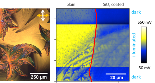
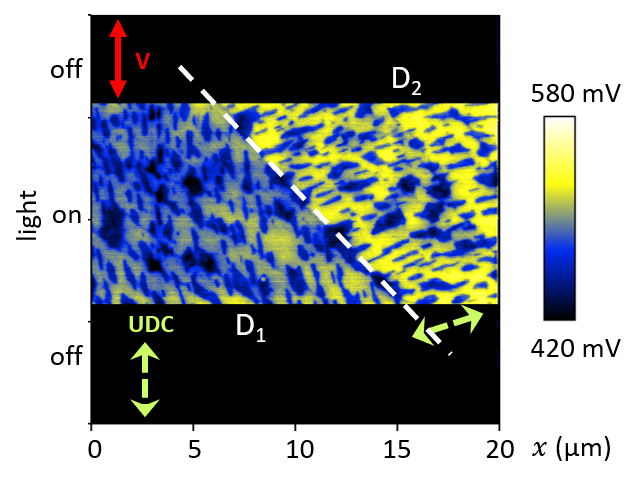
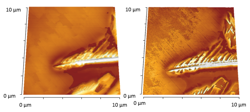
Thin films from anilino-squaraines have been investigated on the nanoscale by scanning probe microscopy. Strong Coulombic coupling leads to polarization dependent absorbance for light. Such films are suitable as active layer in, e.g., bulk-heterojunction photovoltaic cells, operating in the deep red. The surface photovoltage, i.e., the response to linear polarized light in the visible wavelength range was investigated by Kelvin Probe Force Microscopy (KPFM), as shown in the Figure for red light with a wavelength of 700 nm. That way, active parts of the films can be identified on the nanoscale.
- Abdullaeva, O. S.; Balzer, F.; Schulz, M.; Parisi, J.; Lützen, A.; Dedek, K., Schiek, M., Organic Photovoltaic Sensors for Photocapacitive Stimulation of Voltage-Gated Ion Channels in Neuroblastoma Cells, Adv. Funct. Mater. 29, 1805177 (2019)
- Balzer, F.; Abdullaeva, O. S.; Maderitsch, A.; Schulz, M.; Lützen, A., Schiek, M., Nanoscale Polarization-Resolved Surface Photovoltage of a Pleochroic Squaraine Thin Film, Phys. Status Solidi B 257, 1900570 (2020)
Circular Dichroism: From Thin Films to Sensors
In certain chiral squaraine thin films, the circular dichroism is boosted due to a strong excitonic coupling by up to three orders of magnitude compared to similar small organic molecules. This enhancement allows the design of devices sensitive to the circular polarization of light. Based on that, a photodetector has been realized, exceptionally sensitive to the circular polarization of light without any additional optical elements.
- Schulz, M.; Balzer, F.; Scheunemann, D.; Arteaga, O.; Lützen, A.; Meskers, S., Schiek, M., Chiral Excitonic Organic Photodiodes for Direct Detection of Circular Polarized Light, Adv. Funct. Mater. 29, 1900684 (2019)
- Zablocki, J.; Arteaga, O.; Balzer, F.; Hertel, D.; Holstein, J.; Clever, G.; Anhäuser, J.; Puttreddy, R.; Rissanen, K.; Meerholz, K.; Lützen, A., Schiek, M., Polymorphic Chiral Squaraine Crystallites in Textured Thin Films, Chirality 32, 619 (2020)
Stability of Organics at Solid-Liquid Interfaces
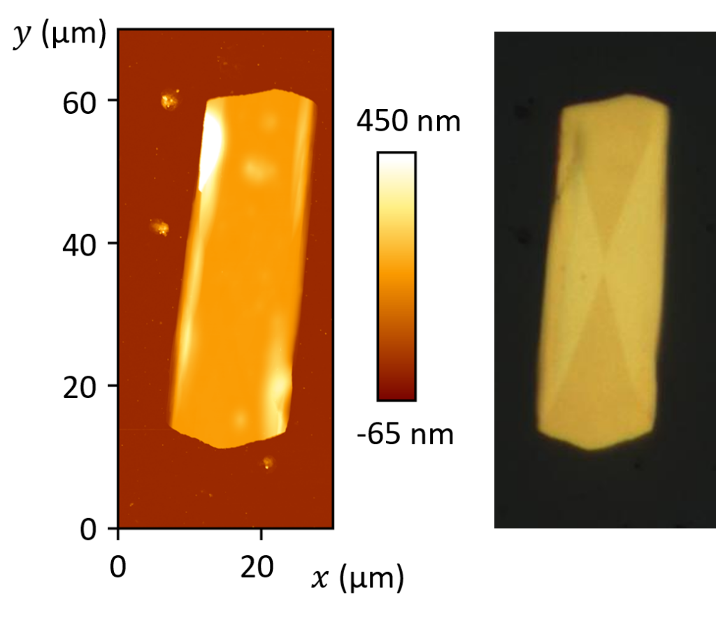
Spending too much time in a water-filled bathtub leads to shriveled fingers. Sugar crystallites are dissolved if one puts them into a cup of coffee. In this project, we try to get a better understanding of structure and stability of organic aggregates and thin films on a microscopic level in a liquid environment parallel to the interaction with white light. In the Figure (left), a topographic microscopy image of a squaraine blend is shown, imaged in air. Different types of aggregates are visible. Immersing these structures in a physiological liquid with simultaneous illumination leads to the dissolution of parts of the structures within a few ten hours, Figure (right). Such investigations on the topography changes but also on the induced electrical properties are of major importance for possible applications such as vision-restoring implants.
- O.S. Abdullaeva, M. Schulz, F. Balzer, J. Parisi, A. Lützen, K. Dedek, M. Schiek, Photoelectrical Stimulation of Neuronal Cells by an Organic Semiconductor-Electrolyte Interface, Langmuir 32 (2016) 8533-8542.
Polarized Light and Organic Aggregates
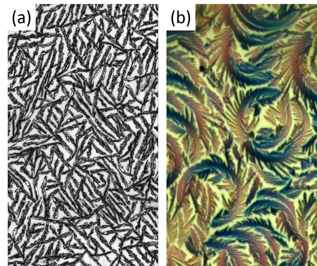
Why are certain beetles colored? Why can you see movies in 3D? Why do certain transparent plastic foils look colored if observed under the blue sky? Why do picture postcards often show deep blue skies and bright white clouds? All these effects are related to the interference and polarization of light. In our labs, thin films of organic molecules are specifically designed and produced to utilize such effects. The image shows microscope images of two typical examples. Like in nature, these ordered structures form by themselves, if the right conditions are provided. The analysis of light’s polarization helps to get a better understanding on how and why these films are formed, how molecules interact within, and how we can improve them for future applications such as clever light detectors.
- F. Balzer, M. Schulz, A. Lützen, M. Schiek, Assembly of Diverse Molecular Aggregates with a Single, Substrate-Directed Molecule Orientation, Soft Matter 12 (2016) 9297-9302.
- F. Balzer, Ch. Röthel, H.-G. Rubahn, A. Lützen, J. Parisi, R. Resel, M. Schiek, Thin film phase and local chirality of surface-bound MOP4 nanofibers, J. Phys. Chem. C 120 (2016) 7653-7661.
Epitaxy of Functionalized Organic Semiconductor Films
A better understanding of the growth of small organic molecules on single crystalline dielectric surfaces is of major interest for the organic electronics community. Contact faces and epitaxy determine, e.g., anisotropic optical and electrical properties of thin films. Therefore, such knowledge is a prerequisite for any device design. Here, we investigate the formation of thin films from a methoxy-functionalized quaterphenylene (MOP4) on single-crystalline KCl and NaCl. MOP4 serves as a model system for a whole class of functionalized para-phenylenes.
In the Figure, results from a number of characterization methods are presented; they deliver complementary results to eventually obtain a complete picture of the growth process. X-ray diffractograms and X-ray pole figures are used to determine the contact planes and the epitaxial orientation of a few hundred nanometer thick films. Low energy electron diffraction (LEED) is able to detect the orientation of only a few nanometer thin films, and to verify the existence of a crystalline wetting layer. Confocal laser scanning microscopy and atomic force microscopy provide the morphology of the sample.
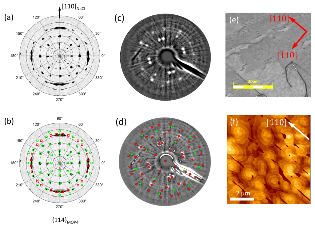
From these methods it is concluded, that the MOP4 molecules vacuum deposited on NaCl and on KCl crystallize into a thin film phase with an increased interplanar spacing compared to the bulk. In-plane diffraction can, however, still be reproduced by the MOP4 bulk lattice constants. Different to other rod-like molecules deposited under similar conditions, the molecules are standing upright on the substrate surface. No wetting layer from lying molecules is found. The contact plane of the MOP4 molecules with the substrates is the (001) plane. Their short unit cell axis is parallel to the substrate <110> and <100> directions. These growth directions are probably driven by an alignment of adsorbate and substrate high-symmetry directions. Note that to some extend also other orientations exist, as documented by, e.g., polarized optical microscopy.
- F. Balzer and M. Schiek, Automated Polarized Microscopy Analysis of Fluorescent and Birefringent Nano- and Microfibers, Chp. 7 in "Bottom-Up Self-Organization in Supramolecular Matter", Springer Series in Materials Science, Vol. 217, 151 - 176.
- F. Balzer, R. Sun, J. Parisi, H.-G. Rubahn, A. Lützen, and M. Schiek, Epitaxial Growth of a Methoxy-Functionalized Quaterphenylene on Alkali-Halide Surfaces, Thin Solid Films 597 (2015) 104 - 111.
Graphene and Graphite as Templates for Epitaxial Growth of Organic Semiconductors
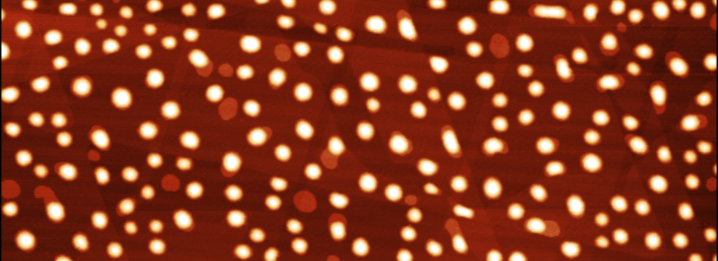
Organic nanowires and nanofibers grown from conjugated molecules such as the para-phenylenes are increasingly used in prototypical devices. Graphene as a growth substrate for organic crystals has interesting perspectives within research and technology. Unlike many other growth substrates, graphene is crystalline, transparent, flat, chemically stable, and electrically conducting, giving it a prospect of being used as combined growth substrate and electrode material. In this project systematic investigations on the optical, morphological, and electrostatic properties of para-phenylene films deposited on graphite and graphene flakes on SiO2/Si are performed. We use atomic force microscopy (AFM) and scanning electron microscopy (SEM) together with polarized-light microscopy (PLM) and X-ray diffraction (XRD) to determine the relative orientation of molecules, epitaxy, and (sometimes surface induced) thin film phases. Kelvin prove force microscopy (KPFM) is used to test the surface potential.
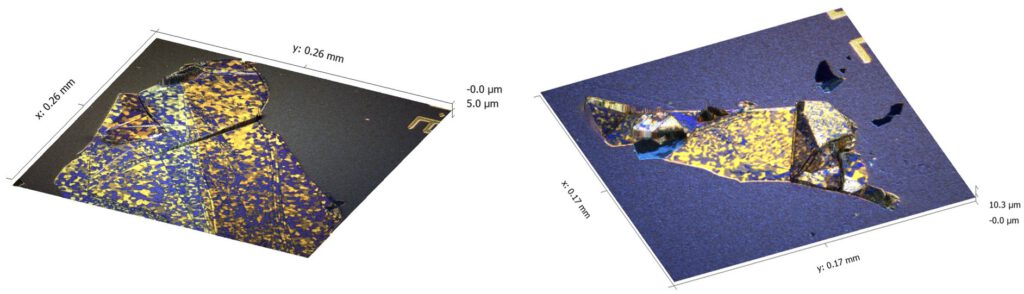
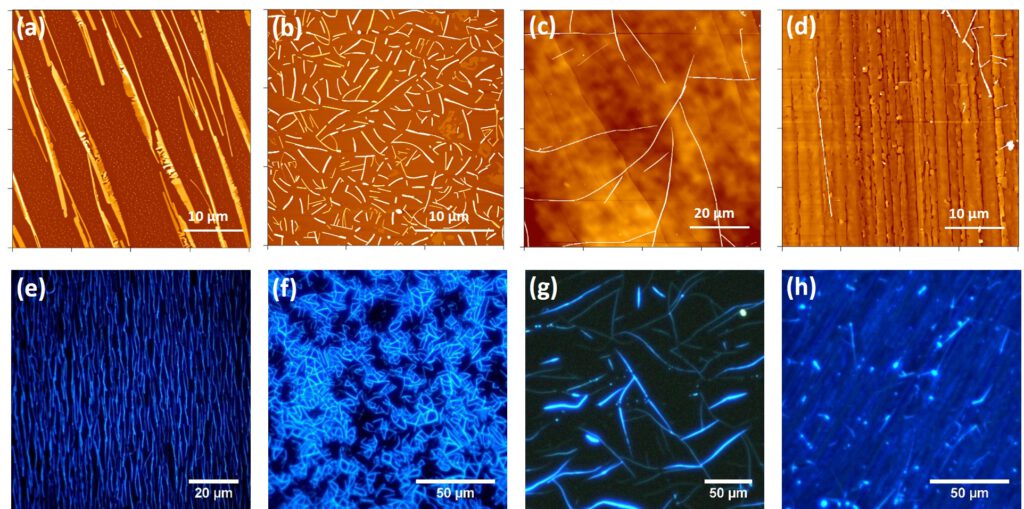
We find that thick (80 nm) p-6P layers form nanostructured crystalline domains on graphite and graphene similar to those found on mica substrates, Fig. 1. Polarized optical microscopy reveals the condensation of two types of fibers on the surface into six domains. White-light bireflectance microscopy can give the same information of molecular orientation as fluorescence microscopy. This is a huge advantage, since UV-bleaching is avoided that way and molecule orientations are used to visualize graphitic domains on the carrier substrate. Ordered p-6P domains are formed even on lithographically processed flakes, having a great potential for technological uses.
The electrostatic properties of different crystal faces of the organic entities are observed with Kelvin probe force microscopy (KPFM); in Fig. 2 this is demonstrated for the growth of a methoxy-functionalized para-phenylene on muscovite mica (a,b) and on graphite (c,d). On muscovite mica only fibers from lying molecules form, having a lower surface potential than the muscovite substrate. On graphite in addition to fibers also islands from upright molecules condense, which show a vastly different surface potential than both the fibers as well as the substrate.

- F. Balzer, H. Henrichsen, M. Klarskov, T. Booth, R. Sun, J. Parisi, M. Schiek, P. Boggild, Directed self-assembled crystalline oligomer domains on graphene and graphite, Nanotechnology 25 (2014) 035602.
- F. Balzer, R. Sun, H.-G. Rubahn, M. Schiek, A. Lützen, Epitaxial growth of a methoxy-functionalized organic semiconductor on dielectric surfaces, Proc. SPIE 8893 (2014) 89830M.
Methoxy Functionalized Nanofibers
A few years ago, methoxy functionalization has shown its potential to improve the formation of organic nanofibers. In this project, we investigate the growth of a methoxy-functionalized para-phenylene (MOP4) on various dielectric substrates as well as the influence of methoxy functionalization on another type of organic semiconductors (naphthyl end-capped oligothiophenes) to shine light on the underlying ordering mechanisms. All molecules have been synthesized in Prof. A. Lützens group, University of Bonn. A combination of optical microscopy (polarized fluorescence and laser-scanning microscopy), birefringence/bireflectance, atomic force microscopy (AFM), low-energy electron diffraction (LEED), and X-ray diffraction (collaboration with Prof. M. Schiek, University of Oldenburg) is used to characterize the growth.
Two different growth modes are observed for the deposition of MOP4 on various dielectric substrates such as muscovite mica, phlogopite mica, graphite, and NaCl; see Fig. 2: the formation of fibers with preferred growth directions, and the epitaxial growth of flat films. We attribute the different behavior to epitaxy and to the decrease of interaction between the molecules and the growth substrate: the interaction between MOP4 and muscovite is strongest, the interaction between MOP4 and sodium chloride weakest. A strong interaction forces the molecules to lie down on the surface, whereas for a weak interaction upright molecules condense. The right choice of the substrate therefore allows the fabrication of organic layers custom tailored for different applications.
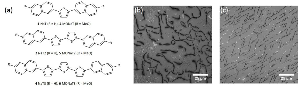
The investigated naphthyl end-capped oligothiophenes are shown in Fig. 2(a). For deposition on muscovite mica methoxy-functionalization improves the fiber growth the same way as for MOP4. Extended focus laser scanning microscope images of NaT and MONaT shown in Figs. 2(b) and (c), respectively, demonstrate the improved crystallization into aligned fibers due to the functionalization. Polarized fluorescence microscopy reveals spatially resolved molecular orientations along the muscovite high-symmetry directions as well as molecule orientations within the aggregates. Different morphological entities (fibers, clusters, flat islands) also show different fluorescence spectra. As many other organic semiconductors, the thionaphthyles samples are not stable in air, but show Ostwald ripening.
- M. Schiek, A. Lützen, R. Koch, K. Al-Shamery, F. Balzer, R. Frese, H.-G. Rubahn, Nanofibers from Functionalized para-Phenylene Molecules, Appl. Phys. Lett. 86 (2005) 153107.
- F. Balzer, M. Schiek, A. Osadnik, I. Wallmann, J. Parisi, H.-G. Rubahn, A. Lützen, Substrate Steered Crystallization of Naphthyl End-Capped Oligothiophenes into Nanowires: The Influence of Methoxy-Functionalization, Phys. Chem. Chem. Phys. 16 (2014) 5747-5754.
- F. Balzer, R. Sun, H.-G. Rubahn, M. Schiek, A. Lützen, Epitaxial growth of a methoxy-functionalized organic semiconductor on dielectric surfaces, Proc. SPIE 8893 (2014) 89830M.